Craniotomy: Correcting Your Problem
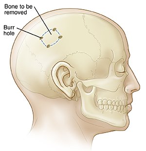
Craniotomy is a surgical opening made in the skull for the treatment of several types of problems in the brain. Special tools are used to remove a piece of the skull and allow access to the brain for surgical treatment. The most common reasons for having a craniotomy include a blood clot (hematoma), tumors, aneurysms, arteriovenous malformations (AVM), and brain abscess.
 |
| The removed bone is also called a bone flap. |
What your surgeon does during your craniotomy depends on your problem. But no matter what, every measure is taken to prevent damage to normal tissue.
-
Brain injury. The source of bleeding is controlled and blood is removed. Damaged tissue may also be cleaned away. The cranial bone (bone flap) might not be put back in place before a few weeks to allow more space for the swollen brain.
-
Brain tumor. As much of the brain tumor as safely possible is removed.
-
Aneurysm. The artery is clipped or sealed at the leak. This prevents more blood from flowing around or into the brain.
-
AVM (arteriovenous malformation). Abnormal arteries and veins are clipped to redirect blood flow to normal vessels and prevent the AVM from leaking blood. After the abnormal arteries and veins are sealed off, they may be removed through the blood vessels using a glue-like substance. This process is called embolization and resection.
Other types of brain procedures
The procedures below may be done alone, or they may be done in addition to a craniotomy. To give access for shunts or stereotactic surgery, burr holes are made in the skull. Your hospital experience before and after these procedures may be about the same as for a craniotomy.
-
Shunts. A shunt is a special type of drain. It is used to decrease pressure on the brain by removing extra cerebrospinal fluid (CSF). The fluid is drained from the brain to the abdomen or another cavity of the body. This is done through a tube that is tunneled under the skin. Once the fluid reaches the abdomen or another cavity, the body absorbs it. Sometimes the CSF drain is connected to an external collection bag for a few days. This is called an external ventricular drain.
-
Brain mapping. The surgeon may keep track of responses during the operation to find regions with important functions.
-
Brain recording and stimulation. This is done as treatment for various conditions, such as epilepsy.
Online Medical Reviewer:
Heather M Trevino BSN RNC
Online Medical Reviewer:
Marianne Fraser MSN RN
Online Medical Reviewer:
Rajadurai Samnishanth
Date Last Reviewed:
3/1/2024
© 2000-2024 The StayWell Company, LLC. All rights reserved. This information is not intended as a substitute for professional medical care. Always follow your healthcare professional's instructions.